External Morphology
Crayfish, being crustaceans, have a body covered by an exoskeleton strongly impregnated with mineral salts. The body is divided into three regions: head (cephalon) (1), thorax (pereion) (2), and abdomen (pleon) (3). The head and thorax are fused into an immovable cephalothorax, the only structural indication of their boundary being the cervical groove (11). The abdomen consists of 6 articulated segments, the last of which ends in the telson (4).
Each body region bears specific appendages:
Each body region bears specific appendages:
- the head appendages are the antennules (5), antennae (6), and mouthparts (one pair of mandibles, two pairs of maxillae, and two pairs of maxillipeds).
- the thoracic appendages (also called pereopods) function primarily in locomotion — walking legs (8). The first pair is modified into chelae (7), used for grasping, digging, and defence.
- the abdominal appendages (also called pleopods) are small and biramous, visible on the ventral surface. Physiologically they generate a water current that oxygenates the gills located at the base of the legs, beneath the thoracic pleurae. During the reproductive season the pleopods are used by the female to carry the eggs, and by the male to transfer spermatophores. In males the first two pairs of pleopods are modified as gonopods. The appendages of the last abdominal segment — in both sexes — are modified into uropods (9), forming the tail fan.

1head (cephalon)
2thorax (pereion)
3abdomen (pleon)
4telson
5antennules
6antennae
7chelae / claws (1st pair of pereopods)
7acarpopodite
7bpropodite
7cdactylopodite
8walking legs (pereopods II–V)
9uropods
10rostrum
11cervical groove
12thoracic pleurae
13abdominal pleurae
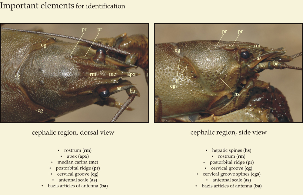
Cephalothorax — identification details
Pârvulescu L. (2010): Crayfish field guide of Romania. Editura Bioflux, Cluj-Napoca, 28 p.
Internal Morphology
The nervous system originates from a cephalic ganglion (1), from which nerves extend to the mouthparts, statocyst, green gland, and eyes, together with a ventral nerve cord (2) that sends branches throughout the entire body, including the appendages. The eyes are compound and mounted on ocular peduncles. The tactile and gustatory senses are highly developed.
The digestive tract begins with the mouth (3), located anteriorly on the ventral face of the cephalothorax and bordered by the mandibles. A short muscular oesophagus leads to the large stomach (4), incompletely divided into two chambers and lined with a complex chitinous endoskeleton. The anterior chamber (cardiac stomach) houses the gastroliths — calcium reserves that supply material for the new cuticle after moulting — while the posterior chamber (pyloric stomach) contains the gastric mill, a series of teeth and ossicles. The intestine (5) is narrow and opens on the ventral face of the telson via the anus (7). The digestive gland — the hepatopancreas (6) — empties into the midgut and is composed of numerous tubules. The intestine, stomach, and oesophagus are all shed and renewed at each moult.
The heart (9) is situated in the posterior part of the cephalothorax and has the form of an elongated sac. Anterior arteries (10) carry blood toward the head, stomach, and hepatopancreas; a posterior artery (11) supplies the abdomen; and an unpaired descending artery (arteria sternalis) connects to the ventral artery (12). The blood is colourless; the plasma contains haemocyanin, a copper-based respiratory pigment (rather than the iron-based haemoglobin found in vertebrates) that turns blue upon contact with air.
The respiratory system is branchial: the gills are situated on the lateral walls of the cephalothorax, attached to the coxae of the pereopods and maxillipeds. The respiratory current is generated by rapid beating of the scaphognathite, an extension of the second maxilla. Excretion is performed by the antennal glands — the green glands (8).
Because the thorax is immovable, it serves as the insertion point for the well-developed abdominal musculature. The appendages also bear strong muscles. Locomotion may occur by forward or backward walking, swimming, or a rapid backward tail-flip (escape response).
The sexes are separate. The gonads (13) lie in the thorax, dorsal to the intestine and ventral to the heart. The openings of the vasa deferentia (14) are located on the coxae of pereopods V in males and III in females. Spermatozoa have an unusual morphology and are grouped into spermatophores, which the male deposits onto the female's sternal plate during mating.

| 1 | cephalic ganglion |
| 2 | ventral nerve cord |
| 3 | mouth |
| 4 | stomach |
| 5 | intestine |
| 6 | hepatopancreas |
| 7 | anus |
| 8 | green gland (antennal gland) |
| 9 | heart |
| 10 | anterior arteries |
| 11 | posterior artery |
| 12 | ventral artery |
| 13 | ovary |
| 14 | oviduct |